


Spirometry
Spirometry with flow volume loops assesses the mechanical properties of the respiratory system by measuring expiratory volumes and flow rates. This test requires the patient to make a maximal inspiratory and expiratory effort. The patient in a sitting position breathes into a mouthpiece, and nose clips are placed to prevent air leak. To obtain interpretable results from spirometry, it is essential that the patient give full effort during testing. At least three tests of acceptable effort are performed to ensure reproducibility of results.
Spirometry is a versatile test of pulmonary physiology. Reversibility of airways obstruction can be assessed with the use of bronchodilators. After spirometry is completed, the patient is given an inhaled bronchodilator and the test is repeated. The purpose of this is to assess whether a patient's pulmonary process is bronchodilator responsive by looking for improvement in the expired volumes and flow rates. In general, a >12% increase in the forced expiratory volume in 1 second (FEV1; an absolute improvement in FEV1 of at least 200 ml) or the forced vital capacity (FVC) after inhaling a beta agonist is considered to be a significant response. However, the lack of an acute bronchodilator effect during spirometry does not exclude a response to long-term therapy.
Similarly, spirometry can be used to detect the bronchial hyperreactivity that characterises asthma. By inhaling increasing concentrations of histamine or methacholine, patients with asthma will demonstrate symptoms and produce spirometric results consistent with airways obstruction at much lower threshold concentration than normals.
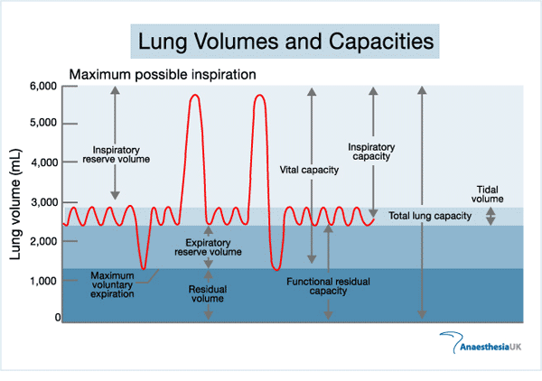
Tidal volume (TV) is the volume of air moved in and out of the respiratory tract (breathed) during each ventilatory cycle.
Inspiratory reserve volume (IRV) is the additional volume of air that can be forcibly inhaled following a normal inspiration. It can be accessed simply by inspiring maximally, to the maximal inspiratory level.
Expiratory reserve volume (ERV) is the additional volume of air that can be forcibly exhaled following a normal expiration. It can be accessed simply by expiring maximally to the maximal expiratory level.
Vital capacity (VC) is the maximal volume of air that can be forcibly exhaled after a maximal inspiration. VC = TV + IRV + ERV.
Residual volume (RV) is that volume of air remaining in the lungs after a maximal expiration. It cannot be expired no matter how vigorous or long the effort. RV = FRC - ERV.
Functional residual capacity (FRC) is the volume of air remaining in the lungs at the end of a normal expiration. FRC = RV + ERV.
Total lung capacity (TLC) is the volume of air in the lungs at the end of a maximal inspiration. TLC = FRC + TV + IRV = VC + RV
Minute volume is the volume of air exhaled per minute.
Maximal breathing capacity (also called "maximal voluntary ventilation") is the maximum volume of air that can be exhaled by voluntary effort in a 15 second interval. This volume is multiplied by 4 and expressed as litres per minute.
Forced expiratory volume 1 (FEV1) - the volume of air that is forcefully exhaled in one second.
Forced vital capacity (FVC) - the volume of air that can be maximally forcefully exhaled.
Ratio of FEV1 to FVC (FEV1/FVC) - expressed as a percentage.
Forced expiratory flow (FEF25 - 75) - the average forced expiratory flow during the mid (25 - 75%) portion of the FVC.
Peak expiratory flow rate (PEFR) - the peak flow rate during expiration.
Spirometry is typically reported in both absolute values and as a predicted percentage of normal. Normal values vary, depending on gender, race, age and height. It is therefore not possible to interpret pulmonary function tests (PFTs) without such information. There is no single set of standard reference values, however, and "normal" varies with the reference value used in each laboratory. It is therefore important to ensure that the reference formulas in a PFT lab are applicable to the patient population being tested.
Forced expiration
Subject inspires maximally then exhales as hard as he or she can.
Volume exhaled in 1 second FEV 1.0
Total volume is the FVC
Normal FEV/FVC ~ 80%
Restrictive (fibrosis) ratio normal or increased
Obstructive (asthma, COAD) usually low

Flow volume loops provide a graphical illustration of a patient's spirometric efforts. Flow is plotted against volume to display a continuous loop from inspiration to expiration. The overall shape of the flow volume loop is important in interpreting spirometric results. The volume versus time curve is a an alternative way of plotting spirometric results and is another useful illustration of patient performance. In normal patients, after a small amount of gas has been exhaled, the flow is limited by airway compression and determined by the elastic recoil of the lung and resistance upstream of that point.
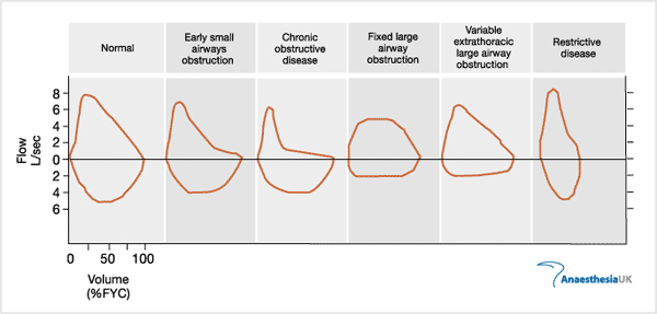
In restrictive diseases, the maximum flow rate is reduced, as is the total volume expired. The flow is abnormally high in the latter part of expiration because of increased recoil.
In obstructive diseases, the flow rate is very low in relation to lung volume, and a scooped out appearance is often seen following the point of maximal flow.
Diffusing capacity (Transfer factor)
The volume of a substance (CO) transferred across the alveoli per minute per unit alveolar partial pressure. CO is rapidly taken up by haemoglobin; its transfer is therefore limited mainly by diffusion. A single breath of 0.3% CO and 10% helium is held for 20 seconds. Expired partial pressure of CO is measured. Normal value 17-25 ml/min/mmHg.
Value is reduced with increased alveolar membrane thickness (e.g. pulmonary fibrosis). May also be reduced with pneumonectomy (results in reduced alveolar membrane).
Multiple breath method
The Rate of N2 washout when the subject is breathing 100% O2
Nitrogen should be reduced by the same fraction with each breath, so that the plot of nitrogen vs the number of breaths produces a straight line. In diseased patients, there is a curved plot from lung units emptying at different rates - there is an initial rapid fall in nitrogen then a slower fall.
Causes of low PaO2
Four causes:
Hypoventilation
Diffusion impairment
Shunt
V/Q mismatch
Raised PaCO2
Two causes:
Hypoventilation
V/Q inequality (does not always cause a high CO2 because the respiratory centre is stimulated to reduce paCO2)
Lung compliance
Volume change per unit change in pressure
Airway resistance
Pressure difference between the alveoli and the mouth per unit of air flow.
Measured with a body plethysmograph. Can also be measured with an oesophageal balloon but tissue viscous resistance is also included.
Closing volume
Another method for detecting early disease is a single breath N2 washout
The subject takes a single VC breath of 100% O2 and the subsequent exhalation of N2 is measured.
Four phases are recognised:
1. Pure dead space is exhaled
2. Dead space and alveolar gas
3. Pure alveolar gas
4. Abrupt increase in N2 concentration
The abrupt increase in phase four signals the closure of airways at the base of the lung and is caused by preferential emptying of the apex, which has a higher concentration of N2. In the apex, it expands less so the N2 is diluted less by the O2. In young people, closing volume is ~ 10% of VC but increases with age to about 40% at 65 years and can enter the FRC so that closure occurs at higher lung volumes.
Control of ventilation
Responsiveness of chemoreceptors and the respiratory centre to CO2 can be measured by having a subject re-breathe into a bag. If CO2 alone is required, then the inspired CO2 should be kept above 200 mmHg. The hypoxic drive can be measured by using a low pO2 but constant pCO2.
Spirometry with flow volume loops assesses the mechanical properties of the respiratory system by measuring expiratory volumes and flow rates. This test requires the patient to make a maximal inspiratory and expiratory effort. The patient in a sitting position breathes into a mouthpiece, and nose clips are placed to prevent air leak. To obtain interpretable results from spirometry, it is essential that the patient give full effort during testing. At least three tests of acceptable effort are performed to ensure reproducibility of results.
Spirometry is a versatile test of pulmonary physiology. Reversibility of airways obstruction can be assessed with the use of bronchodilators. After spirometry is completed, the patient is given an inhaled bronchodilator and the test is repeated. The purpose of this is to assess whether a patient's pulmonary process is bronchodilator responsive by looking for improvement in the expired volumes and flow rates. In general, a >12% increase in the forced expiratory volume in 1 second (FEV1; an absolute improvement in FEV1 of at least 200 ml) or the forced vital capacity (FVC) after inhaling a beta agonist is considered to be a significant response. However, the lack of an acute bronchodilator effect during spirometry does not exclude a response to long-term therapy.
Similarly, spirometry can be used to detect the bronchial hyperreactivity that characterises asthma. By inhaling increasing concentrations of histamine or methacholine, patients with asthma will demonstrate symptoms and produce spirometric results consistent with airways obstruction at much lower threshold concentration than normals.
Tidal volume (TV) is the volume of air moved in and out of the respiratory tract (breathed) during each ventilatory cycle.
Inspiratory reserve volume (IRV) is the additional volume of air that can be forcibly inhaled following a normal inspiration. It can be accessed simply by inspiring maximally, to the maximal inspiratory level.
Expiratory reserve volume (ERV) is the additional volume of air that can be forcibly exhaled following a normal expiration. It can be accessed simply by expiring maximally to the maximal expiratory level.
Vital capacity (VC) is the maximal volume of air that can be forcibly exhaled after a maximal inspiration. VC = TV + IRV + ERV.
Residual volume (RV) is that volume of air remaining in the lungs after a maximal expiration. It cannot be expired no matter how vigorous or long the effort. RV = FRC - ERV.
Functional residual capacity (FRC) is the volume of air remaining in the lungs at the end of a normal expiration. FRC = RV + ERV.
Total lung capacity (TLC) is the volume of air in the lungs at the end of a maximal inspiration. TLC = FRC + TV + IRV = VC + RV
Minute volume is the volume of air exhaled per minute.
Maximal breathing capacity (also called "maximal voluntary ventilation") is the maximum volume of air that can be exhaled by voluntary effort in a 15 second interval. This volume is multiplied by 4 and expressed as litres per minute.
Forced expiratory volume 1 (FEV1) - the volume of air that is forcefully exhaled in one second.
Forced vital capacity (FVC) - the volume of air that can be maximally forcefully exhaled.
Ratio of FEV1 to FVC (FEV1/FVC) - expressed as a percentage.
Forced expiratory flow (FEF25 - 75) - the average forced expiratory flow during the mid (25 - 75%) portion of the FVC.
Peak expiratory flow rate (PEFR) - the peak flow rate during expiration.
Spirometry is typically reported in both absolute values and as a predicted percentage of normal. Normal values vary, depending on gender, race, age and height. It is therefore not possible to interpret pulmonary function tests (PFTs) without such information. There is no single set of standard reference values, however, and "normal" varies with the reference value used in each laboratory. It is therefore important to ensure that the reference formulas in a PFT lab are applicable to the patient population being tested.
Forced expiration
Subject inspires maximally then exhales as hard as he or she can.
Volume exhaled in 1 second FEV 1.0
Total volume is the FVC
Normal FEV/FVC ~ 80%
Restrictive (fibrosis) ratio normal or increased
Obstructive (asthma, COAD) usually low
Flow volume loops provide a graphical illustration of a patient's spirometric efforts. Flow is plotted against volume to display a continuous loop from inspiration to expiration. The overall shape of the flow volume loop is important in interpreting spirometric results. The volume versus time curve is a an alternative way of plotting spirometric results and is another useful illustration of patient performance. In normal patients, after a small amount of gas has been exhaled, the flow is limited by airway compression and determined by the elastic recoil of the lung and resistance upstream of that point.
In restrictive diseases, the maximum flow rate is reduced, as is the total volume expired. The flow is abnormally high in the latter part of expiration because of increased recoil.
In obstructive diseases, the flow rate is very low in relation to lung volume, and a scooped out appearance is often seen following the point of maximal flow.
Diffusing capacity (Transfer factor)
The volume of a substance (CO) transferred across the alveoli per minute per unit alveolar partial pressure. CO is rapidly taken up by haemoglobin; its transfer is therefore limited mainly by diffusion. A single breath of 0.3% CO and 10% helium is held for 20 seconds. Expired partial pressure of CO is measured. Normal value 17-25 ml/min/mmHg.
Value is reduced with increased alveolar membrane thickness (e.g. pulmonary fibrosis). May also be reduced with pneumonectomy (results in reduced alveolar membrane).
Multiple breath method
The Rate of N2 washout when the subject is breathing 100% O2
Nitrogen should be reduced by the same fraction with each breath, so that the plot of nitrogen vs the number of breaths produces a straight line. In diseased patients, there is a curved plot from lung units emptying at different rates - there is an initial rapid fall in nitrogen then a slower fall.
Causes of low PaO2
Four causes:
Hypoventilation
Diffusion impairment
Shunt
V/Q mismatch
Raised PaCO2
Two causes:
Hypoventilation
V/Q inequality (does not always cause a high CO2 because the respiratory centre is stimulated to reduce paCO2)
Lung compliance
Volume change per unit change in pressure
Airway resistance
Pressure difference between the alveoli and the mouth per unit of air flow.
Measured with a body plethysmograph. Can also be measured with an oesophageal balloon but tissue viscous resistance is also included.
Closing volume
Another method for detecting early disease is a single breath N2 washout
The subject takes a single VC breath of 100% O2 and the subsequent exhalation of N2 is measured.
Four phases are recognised:
1. Pure dead space is exhaled
2. Dead space and alveolar gas
3. Pure alveolar gas
4. Abrupt increase in N2 concentration
The abrupt increase in phase four signals the closure of airways at the base of the lung and is caused by preferential emptying of the apex, which has a higher concentration of N2. In the apex, it expands less so the N2 is diluted less by the O2. In young people, closing volume is ~ 10% of VC but increases with age to about 40% at 65 years and can enter the FRC so that closure occurs at higher lung volumes.
Control of ventilation
Responsiveness of chemoreceptors and the respiratory centre to CO2 can be measured by having a subject re-breathe into a bag. If CO2 alone is required, then the inspired CO2 should be kept above 200 mmHg. The hypoxic drive can be measured by using a low pO2 but constant pCO2.


» ultrasound of lung in critical care patients
» respiratory diseases , anaesthetic management 2023
» thoracic anaesthesia 2023
» indication to SICU admission
» electrolyte disturbances
» fluid physiology
» TAP block USG
» Assisted ventilation for surgical patients
» nutrition in critical ill patient
» US workshop in Mosul
» Basics of ultrasound
» الملتقى العلمي الاول للجمعية العراقية للتخدير والعناية المركزة والحد من الالم فرع الموصل
» Geriatric anaesthesia
» thoracic anaesthesia 2022
» chronic pain management
» anaesthesia in respiratory diseases
» anaesthesia for plastic procedures
» ECG for candidate
» postoperative care